A Swiss Army Knife for Neuroscience
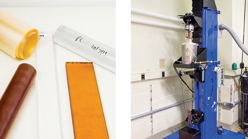
Right: The preform is loaded into this 12-foot-tall fiber-drawing tower.
Various powerful new tools for exploring and manipulating the brain have been developed over the last few years. Some use electronics, while others use light or chemicals.
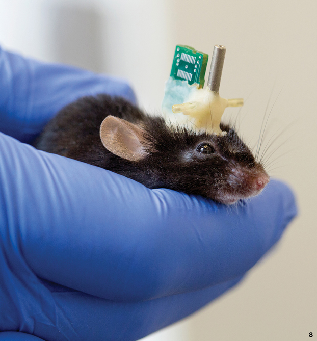
At one MIT lab, materials scientist Polina Anikeeva has hit on a way to manufacture what amounts to a brain-science Swiss Army knife. The neural probes she builds carry light while collecting and transmitting electricity, and they also have tiny channels through which to pump drugs.
That’s an advance over metal wires or silicon electrodes conventionally used to study neurons. Anikeeva makes the probes by assembling polymers and metals into large-scale blocks, or preforms, and then stretching them into flexible, ultrathin fibers.

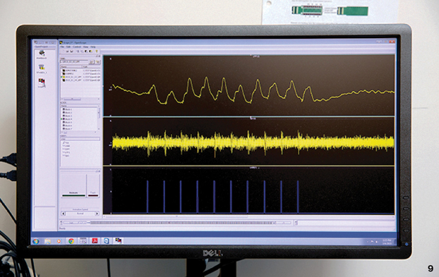
Multifunctional fibers offer new ways to study animal behavior, since they can record from neurons as well as stimulating them. New types of medical technology could also result. Imagine, as Anikeeva does, bionic wiring that bridges a spinal-cord injury, collecting electrical signals from the brain and transmitting them to the muscles of a paralyzed hand.
Anikeeva made her first multifunctional probe while studying at Stanford. It was crude: she simply wrapped metal wires around a glass filament. But this made it possible to combine standard electrode measurements with a new technology, optogenetics, in which light is fired at neurons to activate them or shut them down.
Now Anikeeva, a professor of materials science and engineering, makes probes using a fiber-drawing technology developed by another MIT researcher, Yoel Fink. It’s based on the way silica is heated and pulled to form telecommunications fiber. But it works at lower temperatures, at which many useful polymers become soft enough to stretch.
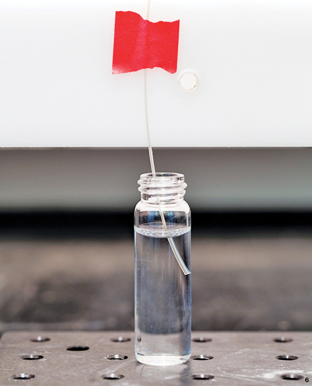
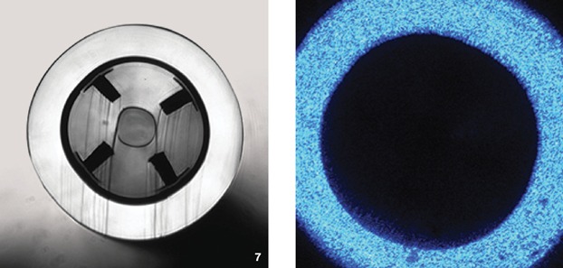
Polymer fibers have a couple of important advantages. One is that they are flexible and mimic the physical properties of tissue. That could allow them to work longer than the stiff metal electrodes neuroscientists have relied on, permitting long-term studies in animals. The second feature of the fibers is that they can combine many functions. Probes made so far have incorporated as many as 36 microwires, optical waveguides, and hollow channels for carrying medicine. There’s no reason not to incorporate sensors to measure temperature or pressure as well. Inside the body, the right materials and structures might even entice nerves to attach to the fibers, the way bone fuses to a hip implant.
The fiber-drawing process shrinks large patterns into microscopic ones, preserving the details. But there are challenges. The tiny wires and tubes have to be stripped, splayed, and soldered by hand to connect them to components such as a recording device a mouse wears on its head. That’s quite a nightmare, says Andres Canales, a graduate student, who hopes to resolve the problem.
Will polymer bio-wires be what ultimately cures paralysis—say, by ferrying nerve signals across an injured spinal cord? “I think it will be a version of this technology, a more sophisticated version,” says Anikeeva. “At least we are going to pursue this route.”

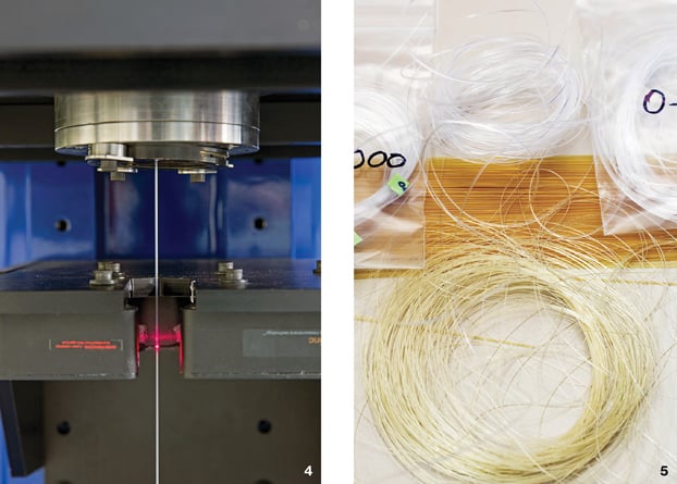
5. Each preform is drawn into as much as one kilometer of fiber. It is now about 1/100th as thick as it was originally.
Keep Reading
Most Popular
Large language models can do jaw-dropping things. But nobody knows exactly why.
And that's a problem. Figuring it out is one of the biggest scientific puzzles of our time and a crucial step towards controlling more powerful future models.
How scientists traced a mysterious covid case back to six toilets
When wastewater surveillance turns into a hunt for a single infected individual, the ethics get tricky.
The problem with plug-in hybrids? Their drivers.
Plug-in hybrids are often sold as a transition to EVs, but new data from Europe shows we’re still underestimating the emissions they produce.
Google DeepMind’s new generative model makes Super Mario–like games from scratch
Genie learns how to control games by watching hours and hours of video. It could help train next-gen robots too.
Stay connected
Get the latest updates from
MIT Technology Review
Discover special offers, top stories, upcoming events, and more.